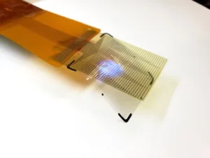
The development of a flexible microdisplay that can monitor and visualize brain activity in real-time during brain surgery has been developed by a collaborative team from the University of California San Diego (UCSD) and Massachusetts General Hospital (MGH),. Itintegrates an electrode grid and LED display to provide a detailed visualization of brain activity, crucial for surgeries involving the removal of brain lesions like tumors and epileptic tissue.

The microdisplay’s key feature is its ability to show different colors to indicate varying brain activities and states. For instance, areas needing surgical removal light up in red, while regions controlling vital functions and thus should be avoided appear in green. This allows neurosurgeons to see and differentiate critical areas in real-time, enhancing surgical precision and safety. The device has already shown its efficacy in animal models, successfully tracking and displaying neural activity that corresponds to specific body areas.
This technology also offers a new way to visualize and manage epileptic seizures. Surgeons can see the onset of a seizure and identify the involved brain nodes, which is crucial for targeted interventions, whether through tissue removal or electrical stimulation to mitigate the seizure.
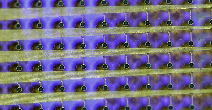
The innovation stems from the extensive research on platinum nanorod electrode grids (PtNRGrids) pioneered by Shadi Dayeh’s lab at UCSD. Dayeh is a professor in the Department of Electrical and Computer Engineering at the UCSD Jacobs School of Engineering and a co-author of the paper. These grids, equipped with LEDs, are capable of recording brain activity at an unprecedented resolution, enabling the visualization of intricate brain functions at a video rate of 40 Hz. The technology’s precision is highlighted by its ability to reduce the standard resection margin in brain surgery from several millimeters to less than one millimeter, potentially leaving less harmful tissue behind.
The microdisplay is crafted using Gallium Nitride which are embedded in a flexible film through a process that includes inkjet printing of quantum dot inks. This technique converts the LEDs’ blue light into multiple colors, allowing for a more nuanced visual representation of neural activity patterns.
Looking ahead, the team plans to develop a microdisplay with up to 100,000 LEDs to achieve a resolution comparable to that of a smartphone screen. This would enable the representation of neural activity with even greater detail, making each LED reflect the activity of fewer neurons, thereby enhancing the clarity and usefulness of the visual data during surgeries. However, the proximity of LED sensors to the PtNRGrids has introduced some interference, a challenge the team is addressing by customizing hardware to refine the device’s performance.
Reference
An electroencephalogram microdisplay to visualize neuronal activity on the brain surface.Sci. Transl. Med.16,eadj7257(2024).DOI:10.1126/scitranslmed.adj7257