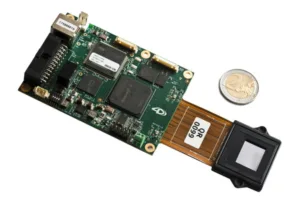
Forth Dimension Displays (ForthDD) , a subsidiary of Kopin Corporation, announced at Photonics West (7 – 12 February 2015) a new QXGA (2048 x 1536) resolution imager and drive board, the QXGA-3DM. The QXGA-3DM combines a high resolution LCoS microdisplay with an application specific drive interface. The system is designed for non-video applications such as the generation of structured light for industrial inspection. The system can be programmed offline, through the RS-232, RS-485 and USB interfaces, and no controller PC is required for normal operation. The drive interface is capable of storing up to 1024 full resolution images. Bi-directional trigger signals ensure accurate synchronization with other system components such as cameras and translation tables.
ForthDD QXGA-3DM LCoS Imager and Drive Board
I was intrigued by the press release statement “the first production shipment of a QXGA-3DM will go to long time ForthDD customer Howard Hughes Medical Institute (HHMI) for use by Nobel Prize for Chemistry 2014 winner Dr. Eric Betzig’s team”. I got in touch with Jim Keeley, Director, Strategic Communications and News at HHMI, to find out exactly how Dr. Betzig’s team was going to use the ForthDD QXGA-3DM.
Mr. Keeley provided me with published material on Dr. Betzig’s use of the previous generation of LCoS imager from ForthDD, the SXGA-3DM with 1280 x 1024 pixels and a fast (<1mS) switching time. This article was published in the 24 October 2014 issue of Science (DOI: 10.1126/science.1257998) and is titled “Lattice Light Sheet Microscopy: Imaging Molecules, Cells, and Embryos at High Spatiotemporal Resolution”.
The ForthDD spatial light modulator (SLM) is used in a binary phase-only mode to generate structured light for an ultra-high resolution imaging system. The imaging system generates 4D movies: x-y-z plus time. The image capture is done at a molecular scale with multiple wavelengths of light in order to image biological processes in living organisms over time.
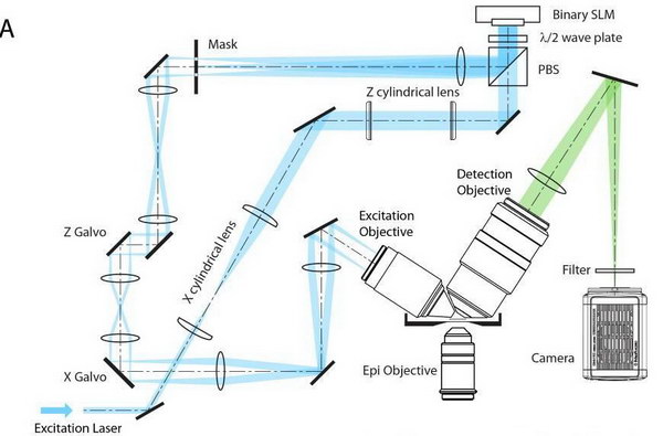 Optical path of the Lattice Light Sheet Microscope (Figure S4A of the Science Article)
According to the article, with lead author Bi-Chang Chen, last author Dr. Betzig and 25 more authors in between, there are other ways to generate structured light for these sorts of imaging systems, including Bessel and confocal illumination. There are two major problems with these prior-art systems, however. First, they cannot generate the structured light on as fine a scale as the SXGA-3DM, reducing the resolution of the system. Second, living biological processes are very sensitive to the illumination light and appear to have a finite tolerence before they are destroyed by the illumination light. In this zero-sum game, if you use an illumination source that generates a higher than necessary illumination intensity, with much of the light wasted, then only shorter, lower resolution movies can be generated before the biological processes are destroyed. Since non-linear, two-photon processes appear to play a part in this destruction, minimizing peak intensity is critical. Presumably, the QXGA-3DM will be able to produce still finer scale structured light with lower intensities, increasing both the spatial and temporal resolution of the system.
According to the paper, “each of the 4D data sets presented here was distilled from tens to hundreds of thousands of raw 2D images acquired from each specimen. This is approximately an order of magnitude more images per specimen than we achieved with linear Bessel beam plane illumination, and nearly two orders of magnitude more than in typical confocal data sets of similar specimens at similar expression levels. Thus, while excitation with a line (Bessel) is better than a point (confocal), a plane (lattice) is better than a line”.
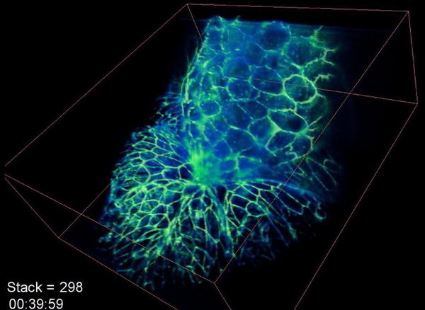
Still from Movie S16 made with a lattice light-sheet microscope at Duke University by one of the Co-Authors
The researchers have posted on Vimeo a number of videos produced by their method. I haven’t had time to look at all of them but according to Jim Keeley 5, 7, S12, S16 are among the best. Keep in mind these are high resolution videos of living organisms at the molecular scale, not computer simulations of these biological processes. For example, S16 shows over 840 time points at 8 sec intervals (112 minutes total time) over a 3D volume of 86 x 80 x 31 μm.
The researchers at HHMI have patented the lattice light-sheet microscopy system. They have licensed it to microscope maker Zeiss and will also share the detailed instructions to researchers who wish to build their own instruments.
Analyst Comment
I find it amazing they can get this level of temporal and spatial resolution in living organisms. I’m sure ForthDD will be happy to sell other research institutions and Zeiss as many SXGA-3DM and QXGA-3DM systems as they need. –Matthew Brennesholtz