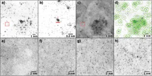
New research out of the Centre for Organic Photonics & Electronics at the University of Queensland in Australia presents a methodological advancement in the characterization of OLED materials. Using high-angle annular dark-field scanning transmission electron microscopy (HAADF-STEM), the authors successfully map the three-dimensional distribution of phosphorescent guest molecules, specifically iridium(III) complexes, within an amorphous host matrix at single atom resolution.
This work is significant for several reasons. Firstly, it addresses a critical aspect of OLED device performance, which hinges on the precise distribution of emissive guest molecules within the host material. Prior to this study, methodologies capable of providing such detailed spatial resolution were limited, leaving a gap in our understanding of how molecular distribution impacts device efficiency and longevity.
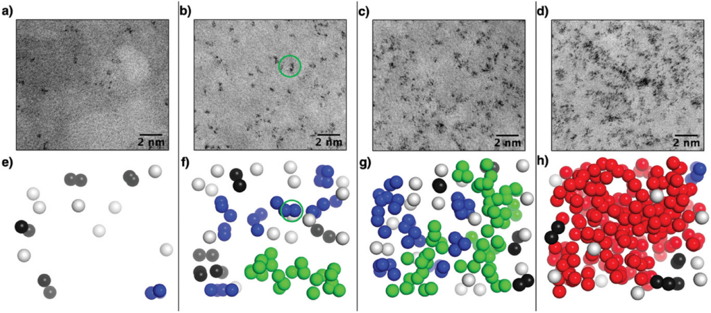
The method developed by the researchers overcomes previous limitations by allowing direct visualization of the guest molecules’ positions. This is achieved by exploiting the contrast between the high electron density of iridium and the surrounding matrix in HAADF-STEM images, facilitating the reconstruction of the guest molecules’ 3D coordinates.
Key findings from this study include the observation that even at low concentrations, iridium(III) complexes tend to cluster rather than distribute uniformly. This clustering behavior becomes more pronounced at higher concentrations, with significant implications for device design and performance. Such insights are crucial for optimizing the balance between luminance, efficiency, and stability in OLED devices.
This research validates results from molecular dynamics simulations of similar systems, providing an empirical basis to theoretical models of guest-host interactions in OLED materials. This validation is essential for the continued development of accurate predictive models for OLED fabrication.
Despite the use of iridium in this study, the techniques and insights gained are broadly applicable. They contribute to the fundamental understanding of material properties in OLEDs and can inform the development of more sustainable, earth-abundant materials that mimic the desirable properties of iridium-based complexes.
Reference
Packman, L., Philippa, B., Pivrikas, A., Burn, P. L., & Gentle, I. R. (2024). Reconstructing the 3D Coordinates of Guest:Host OLED Blends with Single Atom Resolution. Small Methods, 2301305. https://doi.org/10.1002/smtd.202301305