HP has announced plans to offer a version of the company’s Zvr 3D display system that will be targeted for medical applications. This is accomplished by integrating software developed by EchoPixel, Inc. (Mountain View, CA), a maker of 3D medical visualization software that turns diagnostic scans into 3D models, and display hardware developed by zSpace (Sunnyvale, CA).

Two important and related applications in medicine have been identified in which 3D visualization can play an important role.
The first is in medical training. The EchoPixel True 3D Medical Education (t3D/ME) Viewer 1.0 is a software application that “allows medical students to leverage the rich 3D structural anatomy contained in CT and MR images to create a solid foundation of structural 3D biology.”
“t3D/ME provides the advantage of demonstrating complex real anatomy (not drawings) and allowing the student to interactively dissect virtual organs and tissue. t3D/ME enhances the speed with which students understand complex 3D structural relationships, reduces conceptual errors or misunderstandings, and allows a student to repeatedly dissect a virtual 3D body, optimizing cadaver lab assets.”
The second application derives from the fact that current medical visualization techniques have limitations. The problem is that doctors need to mentally integrate a series of 2D images and then consciously think through the data to extract the relevant 3D relationships that define the tissue or organ of interest as well as its neighboring anatomy. The difficulty of this task has the potential of causing a loss of clinically significant information.
The EchoPixel True 3D software enables doctors to experience tissue and organs as apparently real, physical objects making it unnecessary to solve a 3D problem. In this way, doctors are allowed to focus more directly on the immediate clinical problem.
Stating this advantage more specifically, the technology enhances the abilities of doctor to successfully identify an area of interest from inherently 3D medical data sets produced by CT, MRI, ultrasound or any other devices having a 3D modality. By this means, the doctor is assisted in diagnosing ailments, surgical planning and more generally, in using imagery to guide treatment.
Features of the True 3D software that enable real time interaction with the virtual reality system are reported to include:
- Fast super sampling rendering.
- On the fly image processing algorithms (segmentation, registration and etc.)
- Low latency user interaction.
- Real world abstraction user interface.
- No requirement that the data be pre-processed or prepared prior to loading in the system.
In addition, the True 3D protocol provides so-called “Usability tools”. The utility of these tools derive from the fact that “patients are not the same and images are never perfect.” Hence, on-the-fly tools can be used to perform special interactions for identifying, characterizing, sizing and problem solving directly from the raw source data.
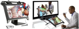
The zSpace 3D display hardware, illustrated on the left in the figure below, creates a stereoscopic image by producing left and right eye images that alternate at 120 Hz. These images are appropriately separated at the user’s eyes by the use of passive glasses (as you’ll see, the system is active, but the glasses passive – Man. Ed.). Further details are as follows.
The zSpace display system uses a twisted nematic LCD to produce the image. This LCD has a 23.6” diagonal and a native resolution of 1920 x 1080. The LED-based backlight located behind the LCD is horizontally segmented. A liquid crystal polarization rotator is located in front of the LCD. It has a corresponding horizontal segmentation. The timing of the imagery presented on the LCD is synchronized with the strobing of the backlight segments and the switching of the polarization rotator segments. An exemplary mode of operation has the right eye images right hand circularly polarized and left eye images left hand circularly polarized. The lenses in the passive glasses are circularly polarized such that the right eye image is transmitted to the user’s right eye and blocked to the user’s left eye and vice versa. By these means, the user is presented with a stereoscopic image.
The head tracking utility in the zSpace system is based on four IR emitters/detectors built into the bezel of the monitor. The light produced by each IR emitter is returned to the adjacent detector by reflection from up to all of the five small retroreflectors embedded in the frame of the passive glasses.
The system also includes a tethered stylus to enable user interaction with the 3D imagery. The stylus combines an IR LED, gyros and accelerometers for tracking, a vibrator to produce haptic effects and three buttons that can be used for user input.
The system can track the user’s head and the stylus over a range that extends 170° horizontally and 160° vertically.
An additional capability of the zSpace system is called zView and is implemented through an optional software/hardware bundle. The hardware includes a camera. This system enables observers to view a 2D representation of the virtual 3D models, the user and the zSpace display on a separate, typically large, conventional display. The zView system is illustrated on the right in the figure below.
A visually impressive video illustrating the EchoPixel medical software running on the zSpace display can be found just below this article.
The Zvr hardware currently listed on the HP web site has a selling price of $3,999. The price of the product when bundled with the EchoPixel medical software is not known at this time nor is the release date. -Arthur Berman
EchoPixel, Ron Schilling, 650-380-2927, [email protected]
zSpace, Dave Chavez, 408-386-1172, [email protected]
Disclosure.
The author has performed paid work for the reported company.