Researchers at the University of Massachusetts Amherst have developed a groundbreaking dual-color optogenetic neural probe capable of both exciting and inhibiting brain activity with different wavelengths of light. The new probe promises unprecedented control over the electrical signals of neurons in the cortex and deep brain regions.
Unlike previous single-color probes limited to either excitation or inhibition, the dual-color design allows enhancing or silencing the same neurons to aid investigation of neural microcircuits. This bidirectional optogenetic capability will enable new insights into diseases like epilepsy and Parkinson’s, according to lead researcher Guangyu Xu.
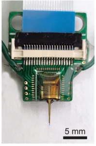
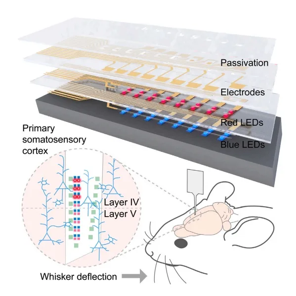
The miniaturized probe integrates closely packed MicroLEDs emitting different colors along with microelectrodes for recording brain activity. Initial tests in mice successfully demonstrated excitation and inhibition of different cortical layers in the somatosensory cortex.
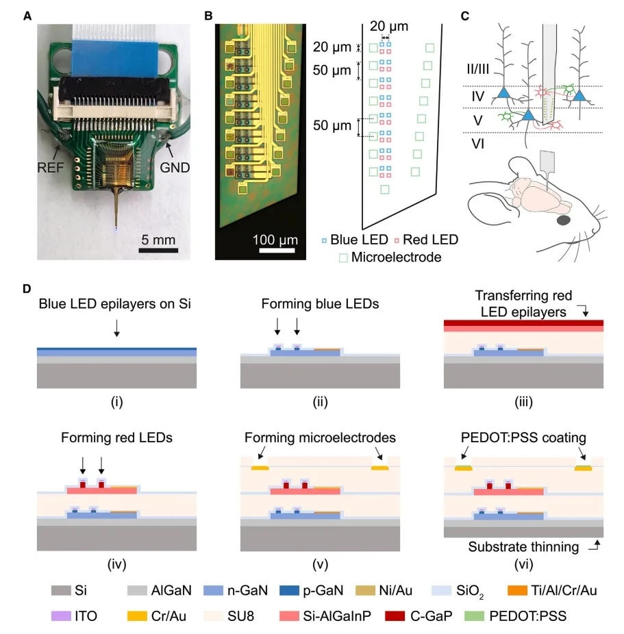
by excitatory (blue triangle) and/or inhibitory (red/green oval) neurons. (D) Simplified fabrication flow. (Source: Cell Reports Physical Sciences)
“We can send one of two colors of light to let neurons in a local brain region become more active or silent, as reflected in neural recording signals,” said lead researcher Guangyu Xu. “This bidirectional capability can lend itself to precisely probing brain circuitry and modeling diseases.”
“Our goal is to keep extending this technology to potentially help map brain circuitry for therapeutic interventions in neurodegenerative diseases,” commented Xu on future research directions. Early studies suggest the probe could eventually be applied to modulate activity in other parts of the central or peripheral nervous systems as well.
Reference
Mao, D., Sun, F., Driscoll, B., Li, Z., & Xu, G. (2023). Close-packed dual-color micro-LEDs enable cortical-layer-specific bidirectional in vivo optogenetic electrophysiology. Cell Reports Physical Science, 4(12), 101702. https://doi.org/10.1016/j.xcrp.2023.101702